Fetal Medicine
Fetal Medicine
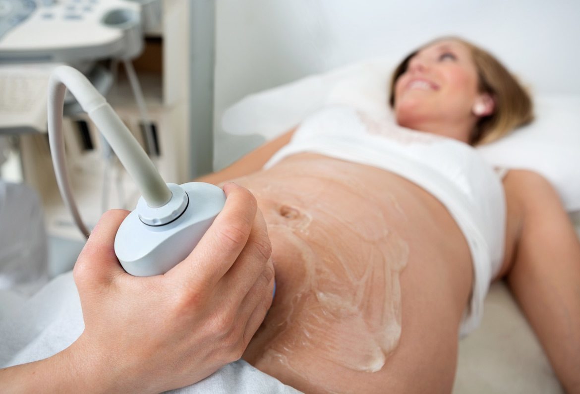
Our Fetal Medicine experts are concerned with the health of the fetus at every stage – monitoring growth & development; predicting, detecting & managing any complications; and treating congenital disorders & anomalies in the womb itself.
WHY FETAL SCANS?
- Evaluate your baby’s growth – monitor baby’s movement, breathing and heart rate.
- Study the placenta and amniotic fluid levels – determine the position of the placenta & assess the amniotic fluid around the baby.
- Identify birth defects & Developmental abnormalities – assess the risk of the baby being affected by certain chromosomal abnormalities e.g. Down syndrome
NT scan
Nuchal translucency (NT) is the sonographic appearance of a collection of fluid under the skin behind the fetal neck in the first-trimester of pregnancy. The term translucency is used, irrespective of whether it is septated or not and whether it is confined to the neck or envelopes the whole fetus. In fetuses with chromosomal abnormalities, cardiac defects and many genetic syndromes the NT thickness is increased.
Screening by NT can detect about 80% of fetuses with trisomy 21 and other major aneuploides for a false positive rate of 5%. The combination of NT and maternal serum free β-hCG and PAPP-A improves the detection to 90%. There is now evidence that the detection rate can increase to about 95% and the false positive rate can be reduced to 3% by also examining the nasal bone, ductus venosus flow and tricuspid flow.
Anomaly Scan
The fetal anomaly scan is a detailed ultrasound scan performed to take a closer look at the womb and the baby between weeks 18-21 of pregnancy.
The Aim
- This is a detailed assessment of the physical structures of the baby at around 18-20 weeks of pregnancy. Special attention is paid to the brain, face, spine, heart, stomach, bowel, kidneys and limbs of the baby.
- Checking the position of the placenta.
- Checking the growth and water around the baby.
- Checking the length of Cervix ( Lower end of uterus) if it is applicable.
The Interpretation
- 2-4% of the babies may have a physical abnormality visible in ultrasonography. As some of the problems may be minor and may not need a serious intervention, except some monitoring, While a few of the other abnormalities may be serious in nature. Based on the nature of the abnormality, it may be lethal, curable or not curable.
- If the placenta is found to be low, a rechecking will be suggested at a later date around 8 months of pregnancy.
- Any problems with the growth and water around the baby or the length of the cervix at this stage will be mentioned and a further action plan will be suggested.
The Preparation and After
- You may eat normally before.
- Usually, we will not need a full bladder for this test.
- Based on the findings, you will be reassured if the baby is found to be normal. If any abnormality is found, Parents will receive full counseling concerning the significance of the abnormality and the various options available.
- You will be given a printed report within 15 minutes in most cases.
Growth Scan And Dopplers
A growth scan is usually done in the last trimester of pregnancy to check for the growth of the fetus along with other parameters like amniotic fluid. The number of growth scans required may vary with each patient depending on the clinical condition of the patient. In general, the first growth scan is done at around 28 weeks. Further scans, if required, are decided based on the findings of this scan. If everything is normal, a second growth scan is usually done around 36 weeks and this scan is usually combined with Doppler scan.
Doppler scans measure the blood flow through the umbilical cord and different parts of the fetal body like the fetal brain and liver. This scan shows whether the fetus is getting all the oxygen and nutrients that it needs via the placenta. This scan is usually combined with the growth scan.
The Information We Get From The Growth Scan Is
- The weight of the fetus, whether it is appropriate, more or less than expected.
- Fluid around the fetus (whether it is adequate, less or more).
- The blood supply going to the fetus.
- Check the level of amniotic fluid.
- Movements of the fetus and well being of the fetus.
- Check the position of the fetus and placenta.
Amniocentesis
At Motherhood, we understand the importance of careful monitoring of the foetus. The Foetal Medicine Department is fully equipped with latest technologies to assess the growth and development of your unborn baby at various stages of the pregnancy. This diagnosis also helps in early detection of illnesses and abnormalities, enabling early treatments and prevent the mother and child from getting harmed.
Foetal medicine services
Necessary preventative and curative treatments are administered in a timely manner at Motherhood hospitals. We are equipped with advanced scanning machines to aid in diagnosis. We have a team of experienced Ultrasound Specialists who monitor both the mother and child with the help of state-of-the-art 3D/4D ultrasound scanning equipment.
First Trimester Scan
- Estimates date of delivery
- Checks pregnancy for twins
- To confirm the site of placental implantation
Second Trimester Scan
- Estimates date of delivery
- Checks organ development
- Assess the amount of amniotic fluid
- Checks blood flow to the baby and within the baby
Third Trimester Scan
- Checks fetal wellbeing
- Tests the baby’s growth rate
- Checks baby’s weight
High-Risk Pregnancy
- Pregnancy with chronic hypertension
- Pregnancy with diabetes
- Pregnancy with heart disease
- Thyroid disorders in pregnancy
- Multiple pregnancies
Genetic Counselling
- Evaluation of family history
- Advice regarding minimally invasive tests (Amniocentesis, CVS, and Fetal Reductions) and non-invasive tests
- Compassionate family counseling
Chorionic Villus Sampling
CVS is a test performed during pregnancy to check for all types of chromosomal abnormalities and prenatal diagnosis of certain genetic disorders like thalassemia in the baby.
Why is it done?
Mostly it is done to rule out chromosomal and genetic disorders in the baby and is offered in the following situations:
- When there is a ‘high risk’ for chromosomal disorders on first trimester screening
- When there is an abnormality in the baby on the first trimester scan
- To rule out genetic disorders like thalassemia, sickle cell disease, muscular dystrophy etc.
- When you have a family history of a genetic disorder for which the genetic mutation is known
How is it done?
CVS is done by passing a needle through the mother’s tummy under local anaesthesia to obtain a small amount of tissue from the placenta (after-birth). The test is done under continuous ultrasound guidance so as to avoid the baby. In some cases, the needle may be needed to put in a second time if enough tissue is not obtained in the first attempt. It is usually done between 11 to 13 weeks of pregnancy; we prefer to do it at around 12 weeks when we can do the first trimester screening as well and rule out any structural abnormality in the baby. The placental tissue is sent to the laboratory for testing. For some tests, both parents blood samples are also needed.
Genetic Testing Counseling
Our fetal medicine fellows will discuss your history, and offer families with pregnancy complications the advanced fetal diagnostic and clinical resources.
We make sure you receive the best possible care.
Evaluation of your family history.
Our genetic counselors will discuss the health of your pregnancy and make recommendations based on your family’s history.
Advice about non-invasive and minimally invasive tests.
When you need help understanding the purpose, risks, benefits and limits of diagnostic procedures, our genetic counselors will talk with you about what to expect.
Compassionate family counseling.
It can be difficult to navigate the emotional challenges that surround the diagnosis of a fetal condition. Our genetic counselors are here to provide you with support when you need it.